ECG.mp4

Images of Human Bone.


Bone fracture : is structural break in normal continuity of a bone. It is usually caused by trauma, example; car accident, fall from height, fall on ground etc.
Types of fracture:
1. Transverse, due to direct trauma
2. Oblique, due to indirect violence
3. Spiral, due to indirect trauma
4. Comminuted, and due to severe compression
5. Green -stick fractures, and
6. Avulsion. In the end of page, there are some images...


Site of the fracture:
1. Intra-articular,
2. Epiphyseal,
3. Metaphyseal, And
4. Diaphyseal.
Fracture may be complete or incomplete.

Displacement:
Deformity of a  bone fractured, it  describes the position of the distal component in the relation the proximal one.
There are six displacement possible below.....


1. Lateral displacement; distal fragment deviates to one side with loss of opposition.
3. Over-riding; distal fragment overlaps the proximal fragment with shortening of the limb.
2. Angulation; loss of normal longitudinal axis of the shaft.
5. Distraction; the fragments are separated by vigorous traction during treatment.
4. Distal fragment is rotated along its long axis, and
7. Depression ; a fragment of bone is displaced.


6. IMPACTION.
Skin damage: According to the condition of the overlying skin fractures are classified into
1. Simple fracture and, in which the skin surface is intact,
2. Compound fracture, in which a laceration in the skin or mucous membrane communicates with fracture hematoma.

Classification of compound fractures shown in the below.

Type of fracture
Skin wound
Soft tissues drainage
Infection risk

Type one
<7 centimeters soft tissue drainage
Minimal Skin wound
<2 % Infection risk
Type two
>7 cm
Moderate Skin Wound
10% Infection risk
Type three
Any size
Severe, example; war wounds or RTA
More than 10%


Case 1.

Over 50 years old female having postmenopausal osteoporosis falls on her right palm of the outstretched  hand in the ground. Patient complaining of pain at wrist, swelling, radial deviation of hand and tenderness in the deformity site.

X-ray is done, shown fracture of distal end of radius of the right hand. And also Dinner-fork deformity of hand.
 X-ray is diagnostic for the colles fracture.

Treatment :
Reduction is done by manipulation under anaesthesia and fixation of fracture by below elbow plaster cast through holding wrist in palmar  flexion and ulnar deviation.

Complications:
1. Malunion of bone,
2. Stiffness of the wrist,
3. Carpal tunnel syndrome, treated by division of flexor retinaculum.
4. Rupture of the extensor pollicis longus tendon and,
5. Sudeck's atrophy.

Case 2:

5 years old boy fallen onto outstretched hand with slightly flexion of the right elbow during playing football.
The boy complaining of pain and swelling in the elbow region, inability to move his right elbow, and  palpable medial and lateral epicondyle.
Patient's radial pulse is felt.

X-ray shows supracondylar fracture of humerus.


Treatment : Depends on type of fracture.

-In the undisplaced fractures and greenstick fractures with angulation less than 20° require no manipulation. Only fixation is done by using a posterior plaster slab and collar and cuff with the elbow flexed for 3 weeks.

- Greenstick fractures with angulation more than 20° reduction by flexion only then fixation as above.
                     While

-Displaced fractures are treated by either..........
1. Closed reduction and fixation by posterior slab  under anaesthesia
2. Closed reduction and percutaneous pinning, and
3. Open reduction and internal fixation if failed closed reduction.
The boy should be admitted to the hospital for observation of the circulation to the hand. If impaired, immediately manage the circulation by removing................     .

Complications of the fracture of the supracondylar of humerus:

1. Nerve injury ;
2. Brachial artery injure ; Volkmann's ischemic contracture.
3. Elbow stiffness and
4. Myositis ossificans.


Case 3:
25 Years old male complaining of inability to raise his left shoulder, and pain due to trauma by car accident since 2012. X- ray is done shown the Anterior Shoulder Dislocation. The patient has epilepsy since 2009.
After management of the shoulder dislocation, patient falls on the ground several times due to epileptic cause as a result several times shoulder dislocation happened as recurrence of shoulder dislocation.

Recurrence should be managed by surgical operations, example;
1. Putti-platt operation ; capsulorrhaphy and shortening of suprascapular muscle.
2. Bankart operation ; reattaching glenoidal labrum to the bony part as shortening the space between...   .
Bankart operation was done for that patient. Some X-ray are  shown in the right corner.
Complications of anterior shoulder dislocation are
1. Axillary nerve injury,
2. Bone fracture,
3. Joint stiffness,
4. Avulsion of supraspinatus tendon and
5. Recurrence.
Some Images below...


Information will be updated later..

Ulnar Claw Hand;

24 Years old M complaining of gradually progressive deformity of left hand after 1.5 years of  surgical operation of left hand due to trauma by rulling machine. Trauma is healed by secondary intention. Operation was done 4 years ago.

Today, patient presents left hand deformity with loss of sensation of the medial part of the palm and fingers (ring and little ) is marked by black ink in the picture.
- On cardboard test: fingers of left hand can not grasp  a  paper sheet due to paralysis of interossei as a result lost of adduction and abduction of fingers.
- Ulnar claw hand due to paralysis of medial two lumbricals due to ulnar nerve injured due to trauma
- Paralysis of adductor pollicis longus; if pinched a paper Between thumb and ring finger , thumb is flexed as froment sign.
- marked atrophy of theinner muscle; muscle wasting. Picture of Claw hand...


Investigation: X-ray is done.
Treatment: conservative treatment and nerve suture as usual.

This patient also have recurrent umbilical hernia.

In this ulnar claw hand case, you need to know the innervation of hand muscles by ulnar nerve and its root.
Muscles are innervated by ulner nerve below.
1. flexor carpi ulnaris**
2. flexor digitorum profundis***
3. lumbrical muscles***
4. opponens digiti minimi
5. flexor digiti minimi
6. abductor digiti minimi
6. interossei** and
7. adductor pollicis**

Root of ulnar nerve: C8 and T1. Shown below


Information will be updated with time.
Ulnar Nerve 


Umbilical Hernia


42 Years old redah complaining of swelling around umbilicus 2 years ago and associated pain 7 days duration.
On examination:
Expansile impulse on cough, doughy sensation, reducible contents, small defect, smooth surface, normal temperature, no tenderness and very small  scar around umbilicus. Smoker; 3 packs per day and lefting heavy objects from 15 years old to 40 years old. No symptoms of other systems. Picture is shown below


Diagnosis : para umbilical hernia.
Operation will be done tomorrow morning.

(Paraumbilical hernia:

A paraumbilical (or umbilical) hernia is a protrusion of the abdominal contents, including mesenteric fat or bowel, through a weak point of the muscles or ligaments near the navel. It can lead to discomfort when fatty tissue gets trapped and a lump can be felt or seen. Whilst they are not usually life-threatening, routine surgical treatment is usually advised to prevent enlargement or strangulation or obstruction of the gut.

Treatment:

The protrusion is put back within the abdomen in the correct position. Stitches are used to strengthen the weakness where the hernia has broken through.

The operation is usually performed under a general anaesthetic.

In most cases this is done as a day case without the need for an overnight stay. From wikipedia ).

Hernia:
Definition
By Mayo Clinic Staff
An umbilical hernia occurs when part of the intestine protrudes through an opening in the abdominal muscles. Umbilical hernia is a common and typically harmless condition. Umbilical hernias are most common in infants, but they can affect adults as well. In an infant, an umbilical hernia may be especially evident when the infant cries, causing the baby's bellybutton to protrude. This is a classic sign of an umbilical hernia.

Many umbilical hernias close on their own by age 1, though some take longer to heal. To prevent complications, umbilical hernias that don't disappear by age 3 or those that appear during adulthood may need surgical repair.

Details about hernia, click the link below:
http://en.m.wikipedia.org/wiki/Hernia


Goitre Egyptian

45 Years old Female  patient complaining of  severe pain in the neck swelling for one week. pain radiating to the ear. Swelling of her neck started 2 years ago. Fine Niddle Aspiration show cancer cells of thyroid tissue. Operation will done today morning.


Clinical pictures:
Local pictures:
1. moderate, regular diffuse thyroid enlargement.
2. soft consistency,
3. Smooth surface,
4. Evidence of high vascularity like systolic expansaile pulsation and thril, bruit maxium at upper poles, and
5. pain radiated or not to ear pathogmonic for cancer, and
6. Up and down with deglutition or not.

Systemic pictures:
1. Intolerance to hot weather,
2. Insomnia,
3. Exophthalmos; True or false,
4. Menorrhagia,
5. Loss of weight, and
6. Jaundice.
7. Diarrhea.

General Examination:
A. Vital signs,
B. Liver enlargement,
C. Upper and lower limbs ; warm hand and hyper reflexia.

Clinical detection of exophthalmos:
1. Exophthalmometer, 13 to 15 mm, more than it ; exophthalmos.
2. Ruler test; if touch the cornea,
3. Naffziger's method; eyeballs protrude beyond the plane of supercillary ridge.
These three are most important test.
4. Russell Frazer 's method ; depth of groove between upper orbital margin and eyeballs is shallow.

Etiology of true exophthalmos: deposition of fat and round cell infiltration in the retrobulbar tissues or may be autoimmune.
and false exophthalmos: retraction of upper eye lids due to contraction of levator palpabrea superior.


Picture is shown  in the right side.

How to diagnose the diffuse toxic goitre and Toxic noduler goitre?
Both of them are diagnosed clinically based on and confirm by investigations.
For diffuse toxic goitre : most intolerance to hot weather where normal people can tolerate, insomnia, exophthalmos  and less arrhythmia. And locally soft consistency, smooth surface,thril and bruit over upper pole of thyroid.
But in the toxic noduler goitre, usually  no exophthalmos but more arrhythmia with other symptoms and locally firm consistency and noduler surface.  Both are due to excessive amount of free  thyroid hormones due to suppression of TSH. Information will be updated with time...............

Treatment:
1. Medical,
2. Radioactive Iodine and,
3. Surgical.
Indications for medical treatment :
- Diffuse toxic goiter
- Preoperative preparation
- Post operative recurrence
- Small gland and
- Refusal of surgery.

Medical treatment includes
- Carbimazole 10 mg three times per day, maximum dose at 60 mg per day, most used drug in Egypt .
It inhibits iodine binding to tyrosine.

- Propylthiouracil 100 mg t.d.s, same action plus prevents peripheral conversation of T4 into T3.
-Propanolol but not used in asthmetic patient, we used Atenolol And
- Iodides.


Inguinal Hernia in Egypt

Day Before yesterday  March 19, 2015, 81 years old diabetic Egyptian patient complaining of bilateral painless swelling in the groin areas and marked  scrotum swelling. Condition started 15 years ago. Patient also has right lower limb edema as a result of cardiac problem and Dyspnea as respiratory problem.
Both swelling increases during cough, visible and palpable but irreducible. Scrotum is full with intestinal contents. Scrotum is hot and red. The case most probably is bilateral indirect inguinal hernia. Shown On the pictures below


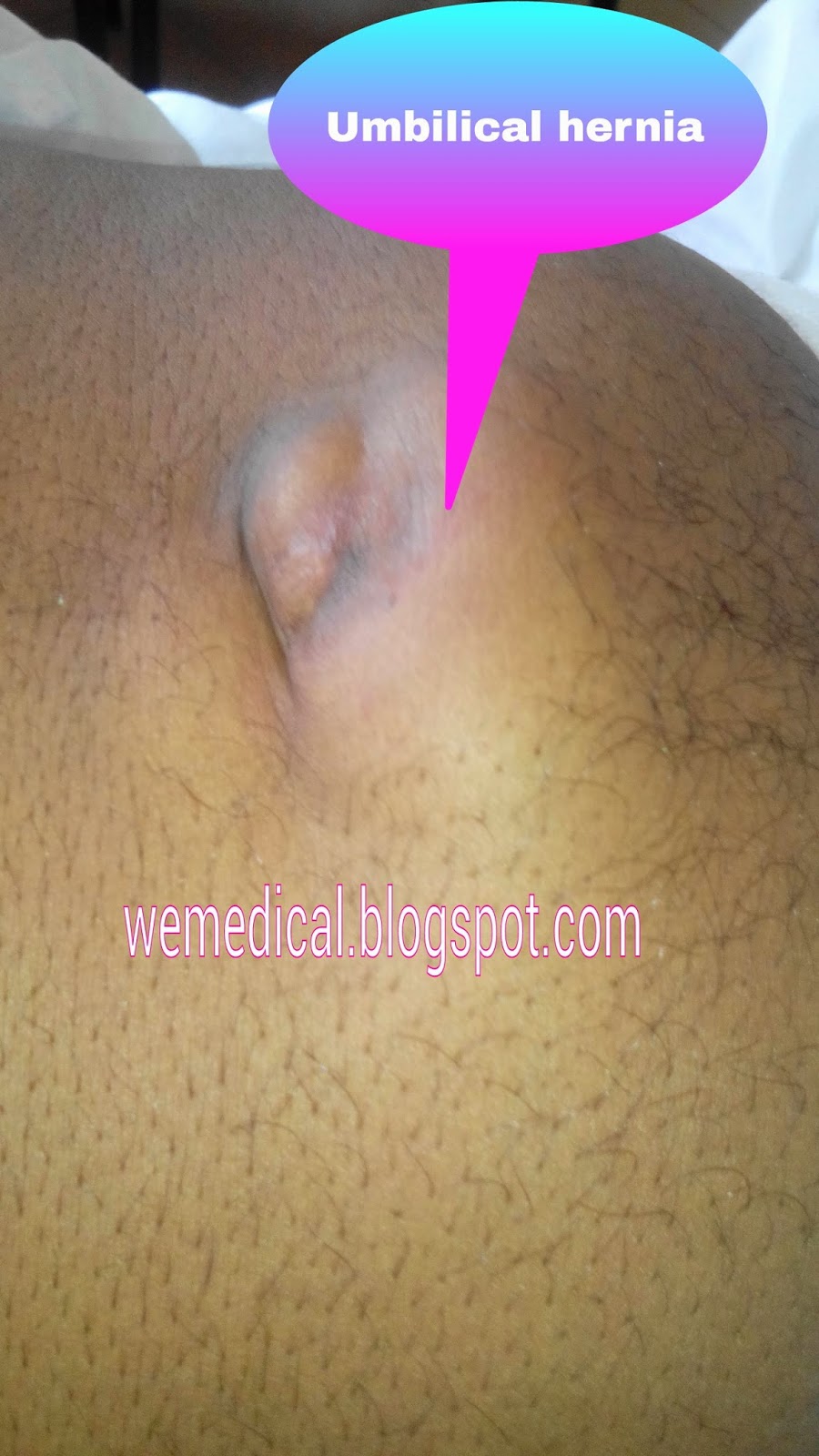
Definition of hernia: it is protrusion of viscera or viscus through a defect into sac characterized by expansile impulse on cough defined as hernia.

On physical examination, the internal ring test revealed no protrusion of contents during cough while ring is obstructed by index finger 1/2 inch above from mid inguinal point. When ring is free, contents come out during cough. The inguinal ring test positive indicates Indirect or oblique inguinal hernia. Characters  of it, during cough contents pass upward, forward, and medially up to scrotum.

External ring test for it: after reduction of contents into abdomen, put your little  finger in the external ring, the ring  very narrow, it just admits your little finger then ask the patient to cough if the contents hit the tip of your finger, it is indirect inguinal hernia but if hit the medial side of your little finger,  it is direct inguinal hernia.
what is internal ring, and why?
It is an opening point to transverse fascia of deep  abdominal muscle, in where testes come out outside the abdomen through that ring like opening. So it forms a canal called inguinal canal. Inguinal canal contains spermatic cords and nerve fibers.

What is mid inguinal point? It is mid way between pubic tubercle and anterior superior iliac spine. 1/2 inch above it, the location of internal ring. Look at pictures down;


Abdominal muscles layers   from superficial to deep in the inguinal area :
1. Skin,
2. Superficial fascia,
3. External oblique muscle,
4. Internal oblique muscle,
5. Transversus abdominis, and
6. Fascia transversalis. Then there is peritoneum. The contents protrude into ring through defect with peritoneum and form the peritoneal pouch within inguinal canal.  Contents could be reduced but peritoneal pouch can not be reduced by clinically. Pictures..


Origins: from iliac crest, inguinal ligament, and ribs.

Insertion: to xiphoid process, linea alba, pubic tubercle and  iliopectineal line. Remember all layers are fleshy fibers except superficial fascia and skin.
 Halfway between pubis symphysis and ASIS is called midpoint of inguinal canal.
A point that is exactly at middle of inguinal ligament is termed 'midpoint of inguinal ligament. 1/2 inch above it, there is a ring called internal ring.

About inguinal canal: it has two openings deep and superior, any circular opening looks like a ring so it is called deep ring but it is in the inguinal area that is why its name is deep inguinal ring or internal ring and it has end second opening at superior is termed superior or external ring or openings.


Boundaries of inguinal canal:
The inguinal canal is made up of:

1. Anterior and posterior walls
2. Superficial and deep rings (openings)
3. Roof and floor (or superior and inferior walls)
We shall go through each component;

The anterior wall is formed by the aponeurosis of the external oblique, and reinforced by the internal oblique muscle laterally.
The posterior wall is formed by the transversalis fascia.
The roof is formed by the transversalis fascia, internal oblique and transversus abdominis.
The floor is formed by the inguinal ligament (a ‘rolled up’ portion of the external oblique aponeurosis) and thickened medially by the lacunar ligament. Look at the picture.

Development :
During periods of increased intra-abdominal pressure, the abdominal viscera are pushed into the inguinal canal. To prevent herniation, the muscles of the anterior and posterior wall contract, and ‘clamp down’ on the canal.

The two openings to the inguinal canal are known as rings. The deep (internal) ring is found above the midpoint of the inguinal ligament. which is lateral to the epigastric vessels. The ring is created by the transversalis fascia, which invaginates to form a covering of the contents of the inguinal canal.

The superficial (external) ring marks the end of the inguinal canal, and lies just superior to the pubic tubercle. It is a triangle shaped opening, formed by the evagination of the external oblique, which forms another covering of the inguinal canal contents. This opening contains intercrural fibres, which run perpendicular to the aponeurosis of the external oblique and prevent the ring from widening.


The inguinal canal is a short passage that extends inferiorly and medially, through the inferior part of the abdominal wall. It is superior and parallel to the inguinal ligament.

It acts as a pathway by which structures can pass from the abdominal wall to the external genitalia.

The inguinal canal also has clinical importance. It is a potential weakness in the abdominal wall, and therefore a common site of herniation.

 Clinically it is important to note that the opening to the inguinal canal is located laterally to the inferior epigastric artery.
Development of the Inguinal Canal
In order to fully comprehend the anatomy of the inguinal canal, we  must first look at its development, and the role the inguinal canal plays in the development of the genitalia. We shall explore the inguinal canal in the context of male development.

 The descent and embryological development of the testes. Note that the processus vaginalis regresses after the descent of the testes
During development, the testes establish in the posterior abdominal wall, and descend into the scrotum. A fibrous cord of tissue called the gubernaculum attaches the inferior portion of the gonad to the future scrotum, and guides them during their descent.

The inguinal canal is the pathway by which the testes are able to leave the abdominal cavity and enter the scrotum. In the embryological stage, the canal is flanked by an outpocketing of the peritoneum, and the abdominal musculature. This outpocketing, the processus vaginalis, normally degenerates, but a failure to do so can result in an indirect inguinal hernia.

In women, there is also a gubernaculum, this attaches the ovaries to the uterus and future labia majora. Because the ovaries are attached to the uterus by the gubernaculum, they are prevented from descending as far as the testes, instead moving into the pelvic cavity. The gubernaculum then becomes the ovarian ligament, and round ligament of uterus.

‘Mid-Inguinal Point’ and ‘Midpoint of the Inguinal Ligament’
These two terms are mentioned frequently in this article, and are often (mistakenly) used interchangeably.

The mid-inguinal point is halfway between the pubic symphysis and the anterior superior iliac spine. The femoral artery crosses into the lower limb at this anatomical landmark.

The midpoint of the inguinal ligament is exactly as the name suggests. The inguinal ligament runs from the pubic tubercle to the anterior superior iliac spine, so the midpoint is halfway between these structures. The opening to the inguinal canal is located just above this point.


The inguinal canal is the pathway by which the testes are able to leave the abdominal cavity and enter the scrotum.
In the embryological stage, the canal is flanked by an outpocketing of the peritoneum, and the abdominal musculature. This outpocketing, the processus vaginalis, normally degenerates, but a failure to do so can result in an indirect inguinal hernia.

In women, there is also a gubernaculum, this attaches the ovaries to the uterus and future labia majora. Because the ovaries are attached to the uterus by the gubernaculum, they are prevented from descending as far as the testes, instead moving into the pelvic cavity. The gubernaculum then becomes the ovarian ligament, and round ligament of uterus.

‘Mid-Inguinal Point’ and ‘Midpoint of the Inguinal Ligament’
These two terms are mentioned frequently in this article, and are often (mistakenly) used interchangeably.

The mid-inguinal point is halfway between the pubic symphysis and the anterior superior iliac spine. The femoral artery crosses into the lower limb at this anatomical landmark.

The midpoint of the inguinal ligament is exactly as the name suggests. The inguinal ligament runs from the pubic tubercle to the anterior superior iliac spine, so the midpoint is halfway between these structures. The opening to the inguinal canal is located just above this point.

Boundaries
The inguinal canal is made up of:

1. Anterior and posterior walls
2. Superficial and deep rings (openings)
3. Roof and floor (or superior and inferior walls)
We shall go through each component..

The anterior wall is formed by the aponeurosis of the external oblique, and reinforced by the internal oblique muscle laterally.
The posterior wall is formed by  transversalis fascia.
The roof is formed by the transversalis fascia, internal oblique and transversus abdominis.
The floor is formed by the inguinal ligament (a ‘rolled up’ portion of the external oblique aponeurosis) and thickened medially by the lacunar ligament.

During periods of increased intra-abdominal pressure, the abdominal viscera are pushed into the inguinal canal. To prevent herniation, the muscles of the anterior and posterior wall contract, and ‘clamp down’ on the canal.

The two openings to the inguinal canal are known as rings. The deep (internal) ring is found above the midpoint of the inguinal ligament. which is lateral to the epigastric vessels. The ring is created by the transversalis fascia, which invaginates to form a covering of the contents of the inguinal canal.

The superficial (external) ring marks the end of the inguinal canal, and lies just superior to the pubic tubercle. It is a triangle shaped opening, formed by the evagination of the external oblique, which forms another covering of the inguinal canal contents. This opening contains intercrural fibres, which run perpendicular to the aponeurosis of the external oblique and prevent the ring from widening.

In men, the spermatic cord passes through the inguinal canal, to supply and drain the testes. In women, the round ligament of uterus traverses through the canal.

The walls of the inguinal canal are usually collapsed around their contents, preventing other structures from potentially entering the canal and becoming stuck.
During periods of increased intra-abdominal pressure, the abdominal viscera are pushed into the inguinal canal. To prevent herniation, the muscles of the anterior and posterior wall contract, and ‘clamp down’ on the canal.

The walls of the inguinal canal are usually collapsed around their contents, preventing other structures from potentially entering the canal and becoming stuck.

Treatment: prevention is the most important, we prevent the risk factors e.g. COPD, smoking, anything that increases intra abdominal pressure.
Mainly surgical, either herniotomy, or hernioplasty, or herniorrhaphy. It depends on........

Main Article: Mesothelioma Most Probably or B. Carcinoma or ILD


43 years old male patient complaining of severe dyspnea on exertion not at rest, Cyanosis on exertion, and dull aching pain over the whole chest. Condition has been started 45 days ago with tolerable dyspnea and high fever, condition was gradually progressive then went to a hospital 15 days later, he was admitted there for 5 days then referred to the Kasr al Ainy hospital on 8 February 2015. Chest X-ray, broncos copy and intercostal tube insertion is done, Result of it are not yet out. But release of fluid through intercostal tube on daily basis showed on the picture.

ILD


38 years old female afaf ala Elam complaining of severe shortness of breath and cough with yellow sputum 10 days ago. Condition started with dyspnea on exertion and cough with small amount of sputum 20 years ago and taking non specific treatment for several time but condition was gradually progressive till 5 years ago. Then with severe dyspnea even at rest, recurrent chest infection, high fever and frequent large amount of sputum, she admitted to Kasr Al Ainy Hospital diagnosed as ILD and she was under medication and oxygen therapy from 5 years ago, but 10 days ago from today February 26 2015 complaining of severe dyspnea at all events, always on sitting with head upward frequent large amount of yellow sputum and chest pain. on physical examination, clubbing of fingers shown on the picture,

 patient on oxygen therapy, crepitation, wheezing, right ankle edema and palpitation.

Patient past history: T.B positive with 9 month anti-TB medication while she was 15 year old and cured with medication. After 3 years later, she had dyspnea and cough as mentioned above and now under oxygen therapy, prednisolone, antibiotic (azathioprine), acetyl cysteine, calcium and folic acid, also with Viagra, the patient under investigation. To gain knowledge of this case read below article.

How does ILD present above symptoms?
what is ILD?
What is the relation between symptoms and ILD?
how to suspect the case of ILD?

ILD: Interstitial Lung Disease means disease in the interstitium of the lung. Interstitium include parenchymal tissue, epithelium, capillary endothelium, basement membrane, perivascular and....vascular lymphatic tissue. In this case, parenchymal tissue is replaced by fibrous tissue through scarring and damaged due to unknown etiology. But suspected cause in this case are TB, asbestos;patient was working in the  cloths factory and bacterial infections due to yellow sputum. Fibrosis of tissues leads to decrease gas exchange as a result dyspnea on exertion, cough and remaining symptoms secondary to infection most probably. Remember, doctors are the brilliant detective for the diseases through taking patient history.
How to suspect or become a detective?
Always from the patient symptoms, every disease have their own symptoms and also every disease have their confirmatory test to confirm the disease, thus you need to memorize these with understanding.
In this case, require the HRCT and  sputum culture with sensitivity test if not respond to treatment.
Chest X-ray may show fine reticular shadow and honeycombing in advanced case of this case.

Treatment: Treat the underlying cause, cause may be autoimmune that is why corticosteroids is the choices but it can not prevent once fibrosis developed. In severe  case, lung transplantation is the best option. Treatment is the same as patient taking medication.


Main Article: T.B


48 years old female live in Faisal, Giza. Complaining of shortness of breath, cough with sputum and chest pain radiating to shoulder in the left side, condition started one year ago with developing the clubbing of her both fingers and pain on both knee, both elbow joints pain with movement 5 month ago for the first time, in the hospital admitted on Thursday, February 22 , 2015. Dyspnea worsens with movement and relieve with rest, sputum more while patient lying on left side with no blood, hands become blue discoloration and palpitation with movement. Lying on right side is normal. The patient had fever at the beginning. Weight loss?, night fever? picture.

Main Article: Bronchiectasis;


34 years old female complaining of cough with large amount of green sputum, shortness of breath associated with paroxysmal nocturnal dyspnea and orthopnea 5 years ago, condition was gradually progressive until 7 days ago that was enough to tolerate for the patient that is why she came to kasr al ainy hospital later with complaining of 13 kg weight loss within 2 months and vomiting, of course also previous symptoms like cough with sputum and dyspnea with clubbing of fingers. Clubbing of fingers is shown on the picture. 

Main Article: Under Attack Egyptian with....


Non diabetic 67 years old patient Mohammad Abdul mannan admitted to Kasr al Ainy hospital two weeks ago and complaining of abdominal distension with mild pain, both lower limbs swelling extended to and above knee; left and right knee respectively, and blurring of vision with yellowish discoloration of sclera of both eyes one month ago. On patient physical examination show: 1. Ascites with shifting dullness, 2. no hepatosplenomegaly felt due to obese patient with marked Ascites, 3. jaundice, and 4. no other sign on psychical examination e.g. dilated veins over abdomen especially around umbilicus and around frank of abdomen. in the first day of admission, administer two pack of blood transfusion, one in the first day and another on second or few days later. Pictures show the sign below.

Main Article: Hemiplegia;


     65 years old hypertensive patient Complaining of sudden weakness of the right side of body and inability to move his right leg and right hand with no fever 7 days ago. Non diabetic, no history of trauma with positive family history of stroke (as I remembered).


Main Article:DKA Patient


     37 years old samy sayed Ex-smoker;20 cigarette  per day for 28 years, works in restaurant , Complaining of disturbed Consciousness Associated with dizziness, nausea, vomiting, weight loss and abdominal pain 4th time with no fever 20 days ago. On physical Examination, hepatomegaly with no dilated vein, no scar, no splenomegaly, no jaundice and reveal loss of sensation of both dorsum of the foot only, no other system are affected. From the history of patient, when he  was 9 years old went to hospital due to his tiredness, week, and polyuria for the first time and blood test shown sugar around 500 mg/dl and prescribed insulin 3 times per day than after one year he lost consciousness than after two years again than.... 4th time with association .

Main Article: Human War against Diseases always in Medicine


When something is made by human that is fully controlled by human due to full knowledge about what has been made. So who knows about human anatomical structures and function of human body??? Main point is we are the peoples or human being, we never create each others. We have knowledge about human being but not full knowledge in every part of human body that is why many diseases are not curable just only controllable e.g.  some autoimmune diseases ,DM etc. human have powerful knowledge than any other creations in the world that is why we human can protect our self from other  harmful creations like pathogens who attack human although human have Defenders inside human body like  very strong `immunity` as a human body defender . Very easy example is everybody knows about windows 8 Defender as it defends windows from any kind of viruses or any kind of harm full entry that may crash the windows.  Human War against Diseases always in Medicine. Human cannot create a life so they have lack of knowledge about that is why many diseases are controllable not curable. Once we human have full knowledge about, we control fully or protect fully from Diseases.


Scleroderma Case;

72 years old male patient complained of symptoms of Myocardial Infarction 7 years ago, today he is complaining of Skin tightening over the hands symmetrically Proximal to distal, difficulty swallowing  and paraesthesia in the hands only.    There is  no kidney failure , no lung affection, and no other system affected

.
Main etiology of this case:

Case:SLE


A new case:
24 years old hypertensive Girl complaining of fatigue, dizziness, paroxysmal nocturnal dyspnea, palpitation, joint pain, photosensitivity, oral ulcer, hands deformities, hair loss and weight loss 15 to 20 kg within 6 months with past history of taking aspirin and ketofen due to her headache from 16 years old till 24 years. In the first time, with two complain of fatigue and dizziness has been treated with folic acid, iron and calcium but no improvement then CBC was done revealed hemoglobin 4mg/dl and high creatinine level. Patient was receiving blood transfusion from being anemic. Today the patient is under investigation to find out renal problem.
25 year old female complaining of 'syncopal


coma' two times, having butter fly skin rash in the check sparing nasolabial fold , gangreen of big toe and anemia , no other menifestations are seen.

allprojects { repositories { google() jcenter() } } dependencies { implementation fileTree(dir: 'libs', include: ['*.jar']) implementation 'com.android.support:appcompat-v7:26.1.0' implementation 'com.google.android.gms:play-services-ads:17.1.1' }